PalkovitsM+2-1972
Notes about [PalkovitsM+2-1972] 1.
This paper give the ratio between mossy fibers, climbing fibers and purkinje cells in the cat cerebellum and a circuit diagram showing the connections.
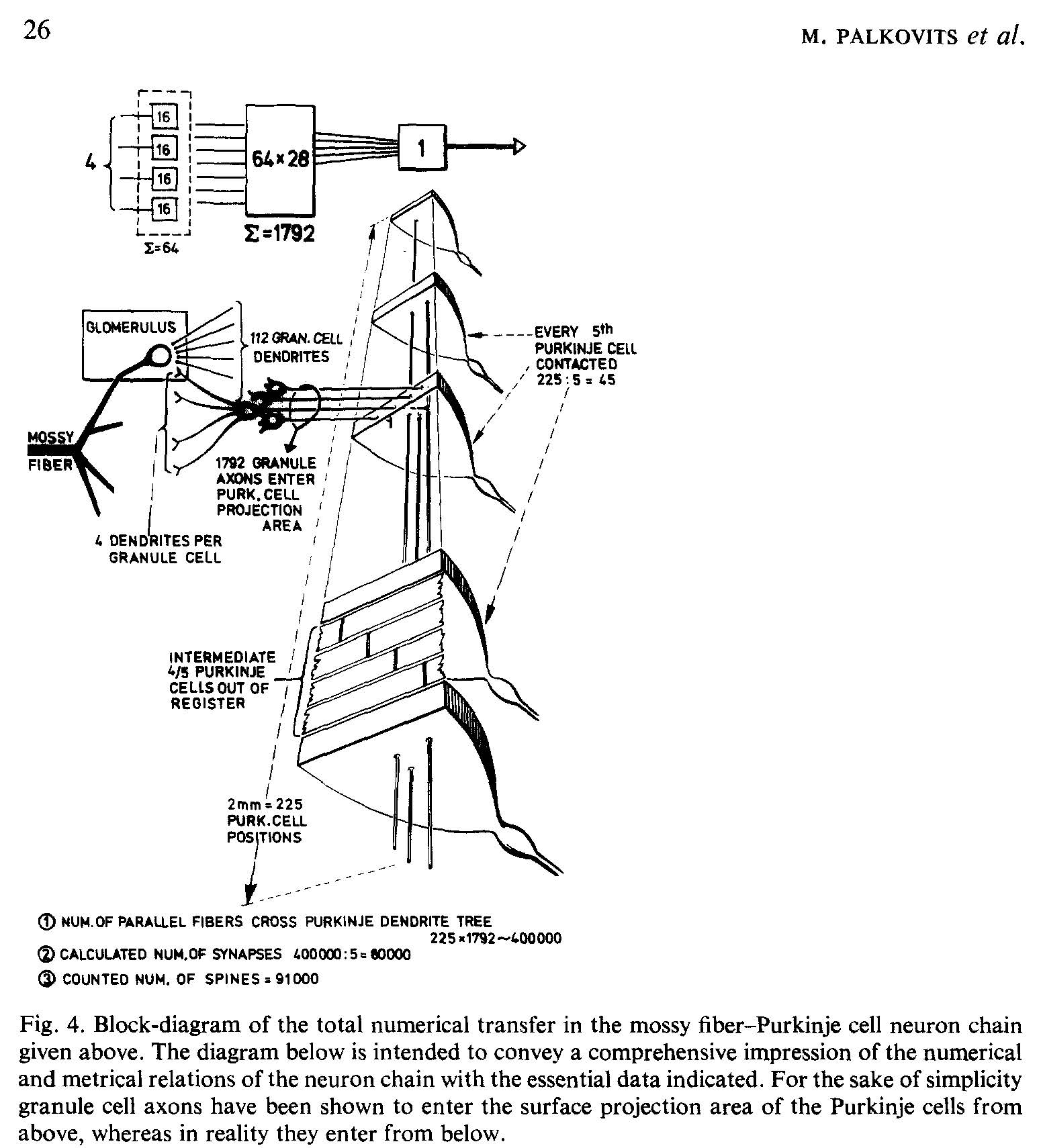
Figure 4 in PalkovitsM+2-1972 [PalkovitsM+2-1972].
“The mossy fiber-Purkinje cell ratio within the folium is thus 4:1.”
From summary:
About 96,000 fibers/sq.mm cross-sectional area enter through the medullary lamina at the base of cerebellar folia in the adult cat. Of these one-sixth are Purkinje axons, one-sixth climbing fibers and the remaining four-sixths of the total number of fibers crossing the base can be assumed to be mossy fibers. The mossy fiber-Purkinje cell ratio within the folium is thus 4:1.
In 1 cu.mm of the granular layer 98,800 glomeruli are found on average (calculated for the living state) in homogeneous distribution and arranged isotropically in space. One mossy fiber breaks up (within a given folium) into about 16-17 mossy rosettes (glomeruli). According to earlier data [10] the granule cell-glomerulus ratio is 27-28:1, the mossy fiber-granule cell ratio is therefore 1:460. The granule cells have 4.17 dendrites, on average; the average mossy rosette is contacted by 112 granule dendrites. The number of postsynaptic units (dendrite digits) is 10.2/dendrite and 1,142/glomerulus. Numerically, the granule cells belonging to one Purkinje cell (1,792) are capable of transmitting impulses from 4 mossy fibers and their 68 rosettes (glomeruli), while the parallel fibers, being 2 mm long, penetrate the dendrite trees of 225 Purkinje cells. Since they establish synapses with only every fifth of these Purkinje cells, the calculated number of parallel fibers-Purkinje spine synapses would be 80,550/ Purkinje cell 11. This calculated value agrees reasonably well with the counted number of Purkinje cell dendritic spines = 91,600.
On the basis of this study series the numerical transfer model of the mossy fiber- Purkinje cell neuron chain can be constructed according to an all-over input-output ratio of 4:1. Further, the numerical, metrical and topological parameters of the divergence from mossy fibers through granule cells and parallel fibers, as well as those of the convergence at granule cell and Purkinje cell level can be established with reasonable accuracy. Since both the numerical and connectivity aspects of the entire neuron model are consistent with one another, and are based on numerous counts and measurements performed using a variety of independent approaches (either on the whole cerebellum or on representative parts), the connectivity model presented is suitable for realistic computer simulation models of the cerebellar cortex.
- 1
Miklos Palkovits, Pal Magyar, and Janos Szentagothai. Quantitative histological analysis of the cerebellar cortex in the cat. IV. Mossy fiber-purkinje cell numerical transfer. Brain Research, 45(1):15–29, October 1972. URL: https://linkinghub.elsevier.com/retrieve/pii/0006899372902132, doi:10.1016/0006-8993(72)90213-2, Notes: PalkovitsM+2-1972.html (this file).